On This Page:ToggleLocationStructureFunctionWhite Matter vs. Grey MatterAssociated DisordersImproving
On This Page:Toggle
On This Page:
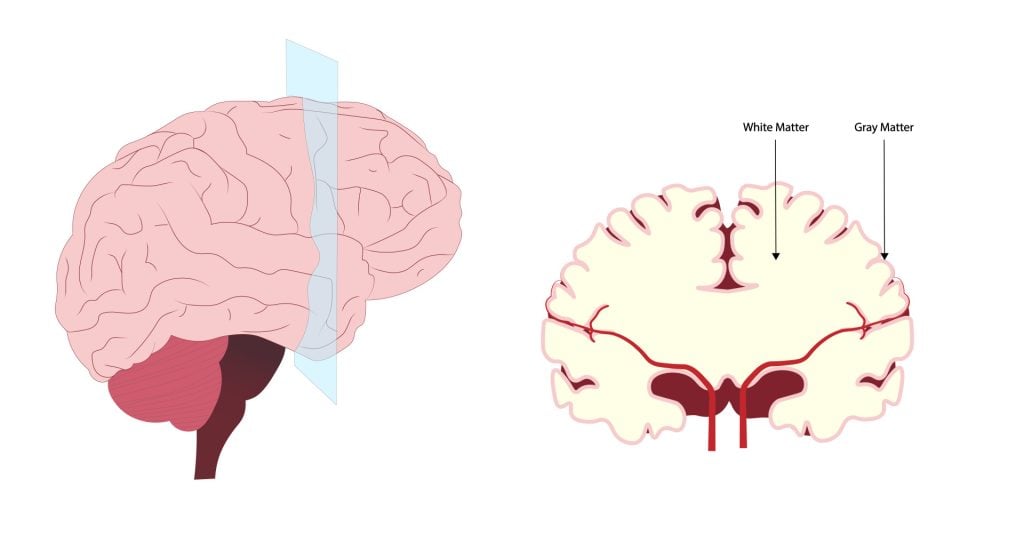
The central nervous system (CNS) is made up of white matter andgrey matter.White matter, comprising about half of the brain, consists of bundles of myelinated axons (nerve fibers).
Located in the deeper parts of the brain, white matter acts as the brain’s communication network, connecting different areas of grey matter and facilitating coordinated brain function.

Where is white matter found?
Its white appearance comes from the myelin sheath, a fatty substance surrounding the axons.
White matter is found in several key areas of the central nervous system:
This distribution allows white matter to effectively connect different regions of grey matter throughout the central nervous system, facilitating communication between various parts of the brain and between the brain and spinal cord.
What White Matter Consists of
White matter consists of millions of bundles of axons, glial cells, and nodes of Ranvier.
Axons are portions of the nerve cell (neuron) that carry electrical signals between different parts of the CNS.
Axons are covered in a fatty insulating substance called myelin sheath.Myelinis white which is what gives white matter its characteristic color.
White matter in the brain and spinal cord is primarily composed of myelinated axons. These axons are nerve fibers that transmit electrical signals between neurons. The “white” appearance comes from the myelin sheath, a fatty insulating layer that surrounds the axons. Myelin not only gives white matter its characteristic color but also increases the speed of neural signal transmission.

These types of cells are known asglial cells, which are cells that provide support or protection to the neurons.
Oligodendrocytes form the myelin and wrap this around the axons up to 150 times. All the layers of myelin are tightly compressed around the axon to ensure it is protected.
On the axon, there are gaps in the myelin sheath called nodes of Ranvier. These unmyelinated gaps cause the impulses traveling along the length of the axon to leap from node to node.
When it does this, the signal increases in velocity so it can reach its destination quicker than traveling down the axon without nodes.
Function
White matter’s primary function is to transmit signals between different brain regions. It therefore plays a crucial role in a variety of brain functions:
White Matter vs. Grey Matter
Below are some of the key differences between white matter and grey matter:
White MatterGrey MatterWhite in colorGrey in colourLocated deep in the brain; outer portion of the spinal cordLocated in outer layer of brain (cortex); central portion of spinal cordComposed mainly of myelinated axons and oligodendrocytesComposed mainly of neuron cell bodies, dendrites, and unmyelinated axonsPrimary function is to transmit signals between different brain regionsPrimary function is to process and analyze informationFast signal transmissionSlower signal transmissionShows structural changes with learning and experienceUndergoes pruning and reorganization during developmentAppears bright on T1-weighted MRI imagesAppears dark on T1-weighted MRI images
Associated Disorders
White matter dysfunction is implicated in various neurological and psychiatric conditions. Multiple sclerosis, a condition where myelin in white matter is destroyed, can lead to motor and sensory disruptions, including fatigue, difficulty walking, and muscle spasms.
In progressive forms, axon damage can result in irreversible neuronal death.
White matter hyperintensities, often referred to as white matter disease, appear as bright white areas on MRI scans due to decreased blood flow. These hyperintensities are associated with a higher risk of stroke and vascular dementia.
Several mental health and developmental conditions have been linked to changes in white matter.
White matter abnormalities might be apparent inautism, post-traumatic stress disorder (PTSD), attention deficit hyperactivity disorder (ADHD), depression, and obsessive-compulsive disorder (OCD).
Schizophrenia has been particularly linked to white matter disruptions across the majority of the brain. This aligns with the typical onset of schizophrenia during adolescence when the forebrain is undergoing myelination
Likewise, studies have found that white matter may play a role in the development of Alzheimer’s disease. Abnormalities in white matter have been found prior to the development of Alzheimer’s.
As well as this, the presence of white matter lesions has been found prior to mild cognitive impairment, a condition that increases the risk for Alzheimer’s disease.
How to Strengthen White Matter
For white matter disease specifically, there is no cure, but there are treatments that can be used to manage the experienced symptoms.
Physical therapy is a common treatment that can help with any balance or walking difficulties someone may develop.
Similarly, managing vascular health could also be an effective way to manage symptoms of white matter disease.
Not smoking and regulating blood pressure (e.g., through blood pressure medications) may help to slow the progression of the disease.
To strengthen white matter, there is support for the use of physical exercise, especially aerobic and weight resistance training.
Physical exercise has also been linked to decreasing the likelihood of developing conditions with cognitive decline, such as dementia and Alzheimer’s.
A suggestion to strengthen white matter could be to pick up a new skill, such as learning to play an instrument or learn a language.
Completing relaxation exercises such as meditation ormindfulness traininghas also been suggested to strengthen white matter functioning.
Finally, methods of protecting the brain from damage are also ways to decrease the risk of white matter dysfunctions.
Avoiding large amounts of alcohol so that balance is not impaired can decrease the risk of falls and damage to the brain and spinal cord.
Also, ensuring a helmet is worn when completing activities such as cycling also minimizes the risk of damage to the brain.
ReferencesFields, R. D. (2010). Neuroscience. Change in the brain’s white matter.Science(New York, NY), 330(6005), 768-769.Klingberg, T., Hedehus, M., Temple, E., Salz, T., Gabrieli, J. D., Moseley, M. E., & Poldrack, R. A. (2000). Microstructure of temporo-parietal white matter as a basis for reading ability: evidence from diffusion tensor magnetic resonance imaging.Neuron, 25(2), 493-500.Newman, T. (2017, August 16).White matter: The brain’s flexible but underrated superhighway. Medical News Todayhttps://www.medicalnewstoday.com/articles/318966Schmahmann, J. D., & Pandya, D. N. (2007). Cerebral white matter—historical evolution of facts and notions concerning the organization of the fiber pathways of the brain. Journal of the History of the Neurosciences, 16(3), 237-267.Schmithorst, V. J., Wilke, M., Dardzinski, B. J., & Holland, S. K. (2005). Cognitive functions correlate with white matter architecture in a normal pediatric population: a diffusion tensor MRI study.Human brain mapping, 26(2), 139-147.Teicher, M. H., Dumont, N. L., Ito, Y., Vaituzis, C., Giedd, J. N., & Andersen, S. L. (2004). Childhood neglect is associated with reduced corpus callosum area.Biological psychiatry, 56(2), 80-85.
References
Fields, R. D. (2010). Neuroscience. Change in the brain’s white matter.Science(New York, NY), 330(6005), 768-769.Klingberg, T., Hedehus, M., Temple, E., Salz, T., Gabrieli, J. D., Moseley, M. E., & Poldrack, R. A. (2000). Microstructure of temporo-parietal white matter as a basis for reading ability: evidence from diffusion tensor magnetic resonance imaging.Neuron, 25(2), 493-500.Newman, T. (2017, August 16).White matter: The brain’s flexible but underrated superhighway. Medical News Todayhttps://www.medicalnewstoday.com/articles/318966Schmahmann, J. D., & Pandya, D. N. (2007). Cerebral white matter—historical evolution of facts and notions concerning the organization of the fiber pathways of the brain. Journal of the History of the Neurosciences, 16(3), 237-267.Schmithorst, V. J., Wilke, M., Dardzinski, B. J., & Holland, S. K. (2005). Cognitive functions correlate with white matter architecture in a normal pediatric population: a diffusion tensor MRI study.Human brain mapping, 26(2), 139-147.Teicher, M. H., Dumont, N. L., Ito, Y., Vaituzis, C., Giedd, J. N., & Andersen, S. L. (2004). Childhood neglect is associated with reduced corpus callosum area.Biological psychiatry, 56(2), 80-85.
Fields, R. D. (2010). Neuroscience. Change in the brain’s white matter.Science(New York, NY), 330(6005), 768-769.
Klingberg, T., Hedehus, M., Temple, E., Salz, T., Gabrieli, J. D., Moseley, M. E., & Poldrack, R. A. (2000). Microstructure of temporo-parietal white matter as a basis for reading ability: evidence from diffusion tensor magnetic resonance imaging.Neuron, 25(2), 493-500.
Newman, T. (2017, August 16).White matter: The brain’s flexible but underrated superhighway. Medical News Todayhttps://www.medicalnewstoday.com/articles/318966
Schmahmann, J. D., & Pandya, D. N. (2007). Cerebral white matter—historical evolution of facts and notions concerning the organization of the fiber pathways of the brain. Journal of the History of the Neurosciences, 16(3), 237-267.
Schmithorst, V. J., Wilke, M., Dardzinski, B. J., & Holland, S. K. (2005). Cognitive functions correlate with white matter architecture in a normal pediatric population: a diffusion tensor MRI study.Human brain mapping, 26(2), 139-147.
Teicher, M. H., Dumont, N. L., Ito, Y., Vaituzis, C., Giedd, J. N., & Andersen, S. L. (2004). Childhood neglect is associated with reduced corpus callosum area.Biological psychiatry, 56(2), 80-85.
![]()
Saul McLeod, PhD
BSc (Hons) Psychology, MRes, PhD, University of Manchester
Saul McLeod, PhD., is a qualified psychology teacher with over 18 years of experience in further and higher education. He has been published in peer-reviewed journals, including the Journal of Clinical Psychology.
Olivia Guy-Evans, MSc
BSc (Hons) Psychology, MSc Psychology of Education
Olivia Guy-Evans is a writer and associate editor for Simply Psychology. She has previously worked in healthcare and educational sectors.
