On This Page:ToggleFunctionsAnatomyLocationThalamus vs. HypothalamusNucleiDamage
On This Page:Toggle
On This Page:
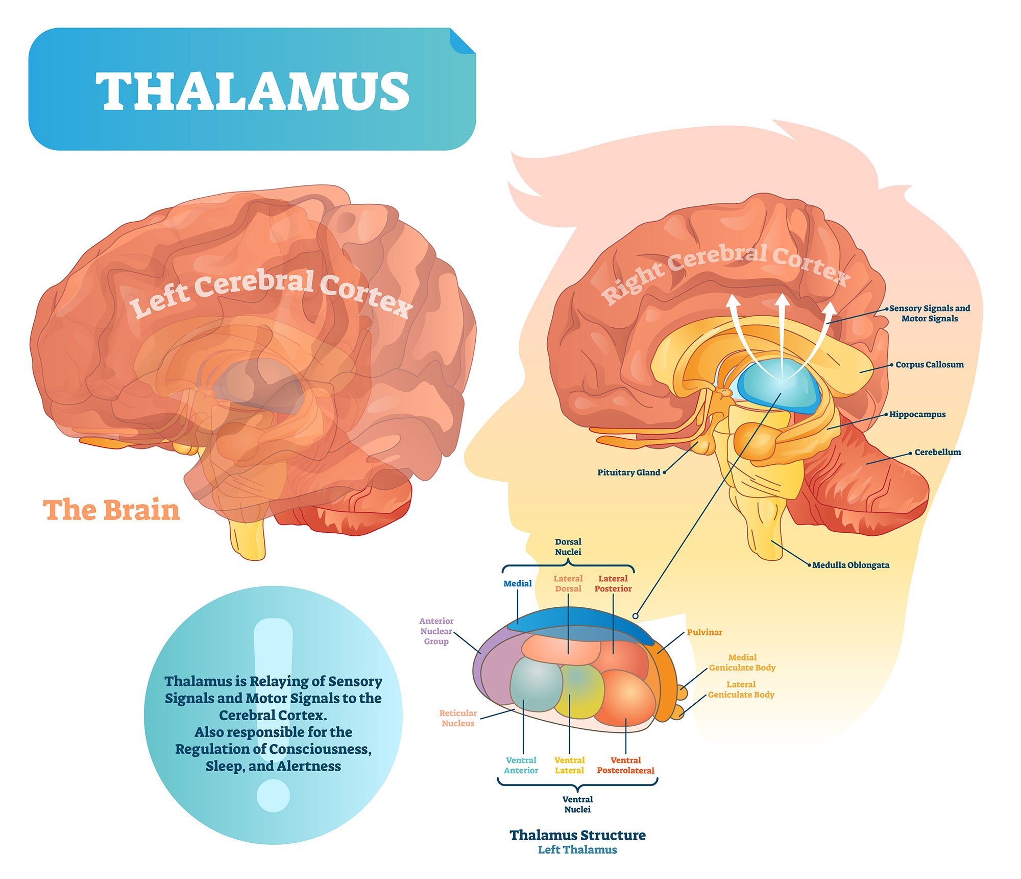
The thalamus is a structure of thebrainthat processes and transmitssensory(except for smell) andmotor informationfrom the body to the cerebral cortex.
The thalamus is often described as thebrain’s relay stationas much of the information that reaches thecerebral cortexfirst stops in the thalamus before being sent to its destination.
Imagine the thalamus is a train station where passengers (or sensory and motor signals) come to catch their train to their intended location (within the brain).
There are two thalami, one in each hemisphere of the brain. They lie above the brain stem and the midbrain (or mesencephalon), which allows for connections of nerve fibers to reach the cerebral cortex in all directions.

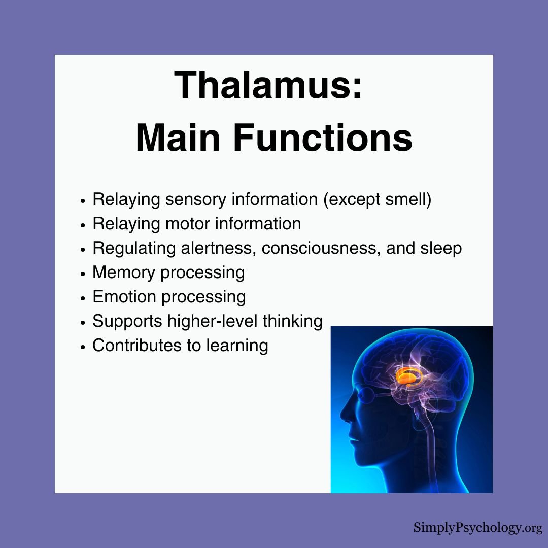
Functions
As the thalamus is heavily involved in relaying informationbetween the cortex and the brain stem, as well as within different cortical structures, it contributes to many brain processes, including:
Sensory Processing & Relay
Motor Signal Control
Attention & Filtering
Consciousness & Arousal
Memory & Cognition
Anatomy
There aretwo thalami, one in each hemisphere of the brain. They are oval-shaped in appearance, almost looking like eggs, withtwo protuberances on the surface.
One of these is known as themedial geniculate bodies, which are important for auditory information processing. The other is thelateral geniculate bodies, which are responsible for the processing of visual sensory inputs.
Location
The thalamus is situatedabove the brainstemin the middle of the brain.
The thalamus is a part of an area called thediencephalon, which includes the hypothalamus, subthalamus, and epithalamus.
Thalamus vs. Hypothalamus
The thalamus and hypothalamus serve distinct functions in the brain.
The thalamus acts as the brain’s relay station, processing and directing sensory and motor signals to the correct areas of the cerebral cortex.
The hypothalamus, located below the thalamus, is thecontrol center for many automatic functionsincluding hormone production, temperature regulation, hunger, thirst, sleep cycles, and emotional responses.
Think of the thalamus as a switchboard operator directing signals, while the hypothalamus is more like the body’s thermostat and control center.
Nuclei within the thalamus
The thalamus is made up ofa series of nuclei, all of which are responsible for the relay of different sensory signals.
The nuclei are bothexcitatory and inhibitoryin nature and receive sensory or motor information from the body, presenting selected information via the nerve fibers to the cerebral cortex.
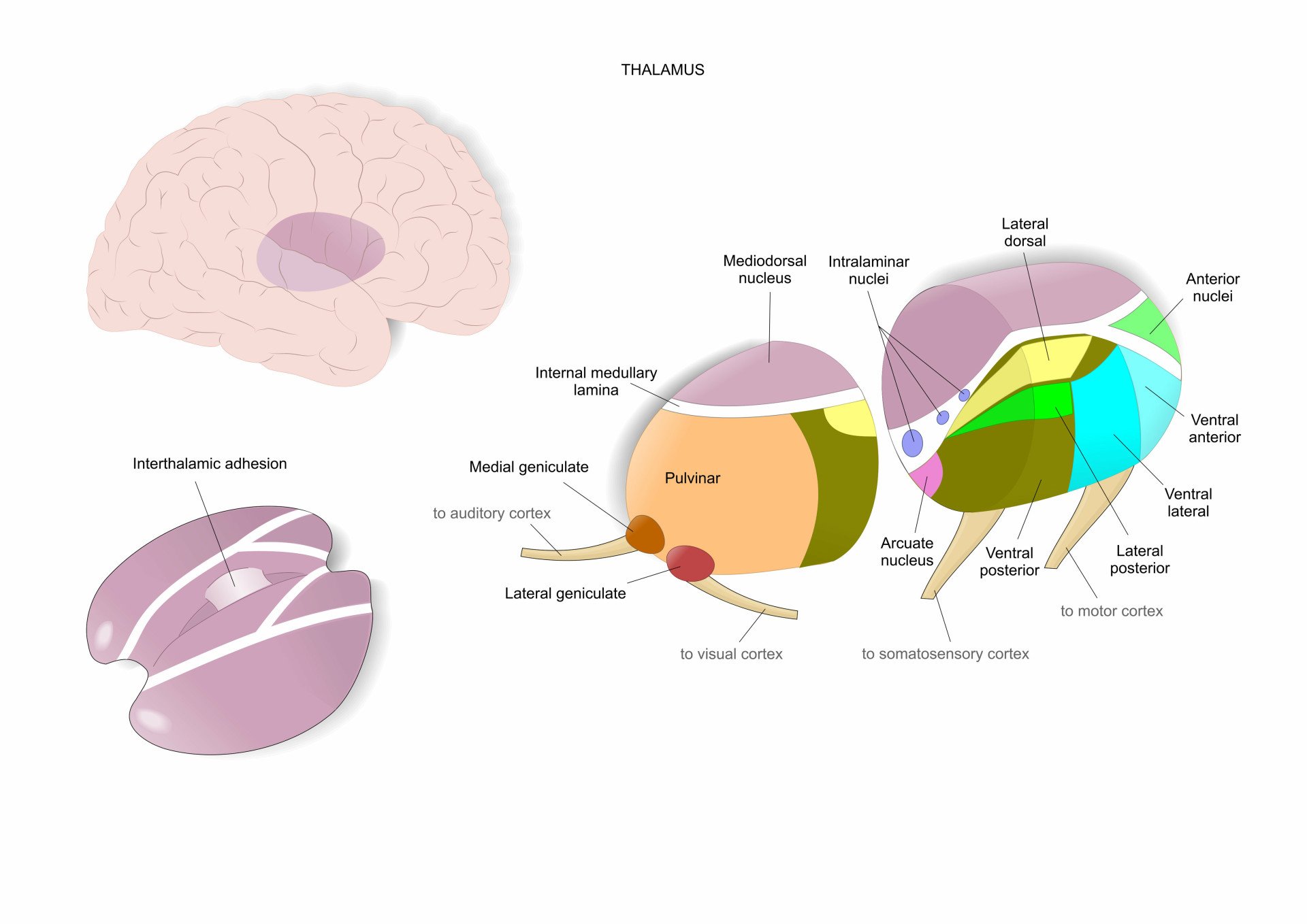
Below are some of the main groups of nuclei within the thalamus and what they are responsible for:
Anterior nucleus
The anterior nucleus is thought to beinvolved with memorydue to its extensive connectivity to the hippocampus.
It is also connected to the mammillothalamic tract (from the mammillary nucleus of the mammillary bodies to the hypothalamus) and the cingulate gyrus (involved in processing emotions and behavior regulation).
As these areas are linked with the limbic system, they are involved in organizing memory and emotion.
Dorsomedial nucleus
The dorsomedial nucleus is thought to be involved inemotional behavior and memory.
This nucleus relays information from theamygdalaand olfactory cortex, which then projects to the prefrontal cortex and the limbic system, in turn relaying them to the prefrontal association cortex.
Because of this, the dorsomedial nucleus has an important role in attention, organization, planning, and higher cognitive thinking.
Ventral posterolateral and ventral posteromedial nucleus
These both act as relay nuclei sending somatosensory information to thesomatosensory cortex, a region thatreceives and processes sensory information about the body.
Further, the ventral posteromedial nucleus receives sensory information regarding the face from the trigeminal nerve.
Ventral anterior and ventrolateral nucleus
These two nuclei are the motor relay nuclei, which receive inputs from thecerebellum and the basal ganglia.
They are thought to be involved in motor functions, and both have pathways leading to the substantia nigra, premotor cortex, reticular formation, and the corpus striatum.
Lateral posterior nucleus
The lateral posterior nucleus is believed to be involved inintegrating sensory input and associating it with cognitive functions.
Its other functions include being able to determine visual stimuli that stands out the most and visually guided behaviors.
Pulvinar nucleus nucleus
The pulvinar nucleus is thought to be involved in theprocessing of visual stimuliand has strong connectivity to the visual cortex.
It projects to the amygdala and the striatum (an area involved in decision-making, reinforcement, and motivation).
This is thought to aid in relaying visual information to guide precise movements as well as relaying visual information to the amygdala.
Medial geniculate and lateral geniculate nucleus
These nuclei are important forrelaying auditory and visual information, respectively.
The lateral geniculate nucleus receives visual information from the retinas of the eyes, which projects to the visual cortex of theoccipital lobe.
The medial geniculate nucleus receives auditory information from the inferior colliculus (a part of the midbrain that is the main auditory center) and projects this to the primary auditory cortex within thetemporal lobe.
Reticular nucleus
The reticular nucleus forms a sheet that makes the outer covering of the thalamus and caninfluence the activity of other nuclei within the thalamus.
The reticular nucleus receives input from the cerebral cortex as well as the dorsal thalamic nuclei.
This is the only nucleus of the thalamus that does not project out to the cerebral cortex but instead modulates the information from other nuclei in the thalamus.
Damage
Since the thalamus is involved in relaying signals from many brain structures, damage to this areacan impact many functions.Symptoms associated with thalamic damage include:
People withschizophreniawere found to have significantly reduced thalamic volume compared to those without schizophrenia (Cosciaet al.,2009).
This reduced size was thought to be associated with weakened neuropsychological functioning and specific difficulties with language, motor, and executive skills.
Lower and weaker functional connectivity in the thalamus was also found inautistic individuals(Tomasi & Volkow, 2019).
‘Non-typical’ thalamic connectivity in temporal and motor areas was also found inautisticindividuals (Woodwardet al.,2017).
ReferencesCoscia, D. M., Narr, K. L., Robinson, D. G., Hamilton, L. S., Sevy, S., Burdick, K. E., Gunduz-Bruce, H., McCormack, J., Bilder, R. M. & Szeszko, P. R. (2009). Volumetric and shape analysis of the thalamus in first‐episode schizophrenia.Human brain mapping, 30(4), 1236-1245.Mandal, A. (2021, February 15).What is the Thalamus?News Medical Life Sciences.https://www.news-medical.net/health/What-is-the-Thalamus.aspx#:~:text=The%20thalamus%20is%20a%20small,signals%20to%20the%20cerebral%20cortexTomasi, D., & Volkow, N. D. (2019). Reduced local and increased long-range functional connectivity of the thalamus in autism spectrum disorder.Cerebral Cortex, 29(2), 573-585.Torrico, T. J., & Munakomi, S. (2019).Neuroanatomy, Thalamus.Woodward, N. D., Giraldo-Chica, M., Rogers, B., & Cascio, C. J. (2017). Thalamocortical dysconnectivity in autism spectrum disorder: an analysis of the autism brain imaging data exchange.Biological Psychiatry: Cognitive Neuroscience and Neuroimaging, 2(1), 76-84.
References
Coscia, D. M., Narr, K. L., Robinson, D. G., Hamilton, L. S., Sevy, S., Burdick, K. E., Gunduz-Bruce, H., McCormack, J., Bilder, R. M. & Szeszko, P. R. (2009). Volumetric and shape analysis of the thalamus in first‐episode schizophrenia.Human brain mapping, 30(4), 1236-1245.
Mandal, A. (2021, February 15).What is the Thalamus?News Medical Life Sciences.https://www.news-medical.net/health/What-is-the-Thalamus.aspx#:~:text=The%20thalamus%20is%20a%20small,signals%20to%20the%20cerebral%20cortex
Tomasi, D., & Volkow, N. D. (2019). Reduced local and increased long-range functional connectivity of the thalamus in autism spectrum disorder.Cerebral Cortex, 29(2), 573-585.
Torrico, T. J., & Munakomi, S. (2019).Neuroanatomy, Thalamus.
Woodward, N. D., Giraldo-Chica, M., Rogers, B., & Cascio, C. J. (2017). Thalamocortical dysconnectivity in autism spectrum disorder: an analysis of the autism brain imaging data exchange.Biological Psychiatry: Cognitive Neuroscience and Neuroimaging, 2(1), 76-84.
![]()
Saul McLeod, PhD
BSc (Hons) Psychology, MRes, PhD, University of Manchester
Saul McLeod, PhD., is a qualified psychology teacher with over 18 years of experience in further and higher education. He has been published in peer-reviewed journals, including the Journal of Clinical Psychology.
Olivia Guy-Evans, MSc
BSc (Hons) Psychology, MSc Psychology of Education
Olivia Guy-Evans is a writer and associate editor for Simply Psychology. She has previously worked in healthcare and educational sectors.
