On This Page:ToggleBrain PartsRight Brain vs. Left BrainLobes of the BrainCerebral CortexDeep StructuresVentricles & Cerebrospinal FluidNeuronsGlial CellsCranial Nerves
On This Page:Toggle
On This Page:
The brain receives information from sensory receptors and sends messages to muscles and glands. It is the center of all conscious awareness and is divided into different lobes with different functions. It contains the cerebrum, about 85% of the total mass.The brain controls all functions of the body, interprets information from the outside world, and defines who we are as individuals and how we experience the world.The brain receives information through our senses: sight, touch, taste, smell, and hearing. This information is processed in the brain, allowing us to give meaning to the input it receives.The brain is part of the central nervous system (CNS) along with the spinal cord. There is also a peripheral nervous system (PNS) comprised of31 pairs of spinal nervesthat branch from the spinal cord and cranial nerves that branch from the brain.Brain PartsThe brain is composed of the cerebrum, cerebellum, and brainstem (Fig. 1).Figure 1.The brain has three main parts: the cerebrum, cerebellum, and brainstem.CerebrumThe cerebrum is the largest and most recognizable part of the brain. It consists ofgrey matter(thecerebral cortex) andwhite matterat the center. The cerebrum is divided into two hemispheres, the left and right, and contains the lobes of the brain (frontal, temporal, parietal, and occipital lobes).The cerebrum produces higher functioning roles such as thinking, learning, memory, language, emotion, movement, and perception.The CerebellumThecerebellumis located under the cerebrum and monitors and regulates motor behaviors, especially automatic movements.This structure is also important for regulating posture and balance and has recently been suggested for being involved in learning and attention.Although the cerebellum only accounts for roughly 10% of the brain’s total weight, this area is thought to contain more neurons (nerve cells) than the rest of the brain combined.BrainstemThe brainstem is located at the base of the brain. This area connects the cerebrum and the cerebellum to the spinal cord, acting as a relay station for these areas.The brainstem regulates automatic functions such as sleep cycles, breathing, body temperature, digestion, coughing, and sneezing.
The brain receives information from sensory receptors and sends messages to muscles and glands. It is the center of all conscious awareness and is divided into different lobes with different functions. It contains the cerebrum, about 85% of the total mass.
The brain controls all functions of the body, interprets information from the outside world, and defines who we are as individuals and how we experience the world.The brain receives information through our senses: sight, touch, taste, smell, and hearing. This information is processed in the brain, allowing us to give meaning to the input it receives.The brain is part of the central nervous system (CNS) along with the spinal cord. There is also a peripheral nervous system (PNS) comprised of31 pairs of spinal nervesthat branch from the spinal cord and cranial nerves that branch from the brain.Brain PartsThe brain is composed of the cerebrum, cerebellum, and brainstem (Fig. 1).Figure 1.The brain has three main parts: the cerebrum, cerebellum, and brainstem.CerebrumThe cerebrum is the largest and most recognizable part of the brain. It consists ofgrey matter(thecerebral cortex) andwhite matterat the center. The cerebrum is divided into two hemispheres, the left and right, and contains the lobes of the brain (frontal, temporal, parietal, and occipital lobes).The cerebrum produces higher functioning roles such as thinking, learning, memory, language, emotion, movement, and perception.The CerebellumThecerebellumis located under the cerebrum and monitors and regulates motor behaviors, especially automatic movements.This structure is also important for regulating posture and balance and has recently been suggested for being involved in learning and attention.Although the cerebellum only accounts for roughly 10% of the brain’s total weight, this area is thought to contain more neurons (nerve cells) than the rest of the brain combined.BrainstemThe brainstem is located at the base of the brain. This area connects the cerebrum and the cerebellum to the spinal cord, acting as a relay station for these areas.The brainstem regulates automatic functions such as sleep cycles, breathing, body temperature, digestion, coughing, and sneezing.
The brain controls all functions of the body, interprets information from the outside world, and defines who we are as individuals and how we experience the world.
The brain receives information through our senses: sight, touch, taste, smell, and hearing. This information is processed in the brain, allowing us to give meaning to the input it receives.
The brain is part of the central nervous system (CNS) along with the spinal cord. There is also a peripheral nervous system (PNS) comprised of31 pairs of spinal nervesthat branch from the spinal cord and cranial nerves that branch from the brain.
Brain Parts
The brain is composed of the cerebrum, cerebellum, and brainstem (Fig. 1).

Figure 1.The brain has three main parts: the cerebrum, cerebellum, and brainstem.
Cerebrum
The cerebrum is the largest and most recognizable part of the brain. It consists ofgrey matter(thecerebral cortex) andwhite matterat the center. The cerebrum is divided into two hemispheres, the left and right, and contains the lobes of the brain (frontal, temporal, parietal, and occipital lobes).
The cerebrum produces higher functioning roles such as thinking, learning, memory, language, emotion, movement, and perception.
The Cerebellum
Thecerebellumis located under the cerebrum and monitors and regulates motor behaviors, especially automatic movements.
This structure is also important for regulating posture and balance and has recently been suggested for being involved in learning and attention.
Although the cerebellum only accounts for roughly 10% of the brain’s total weight, this area is thought to contain more neurons (nerve cells) than the rest of the brain combined.
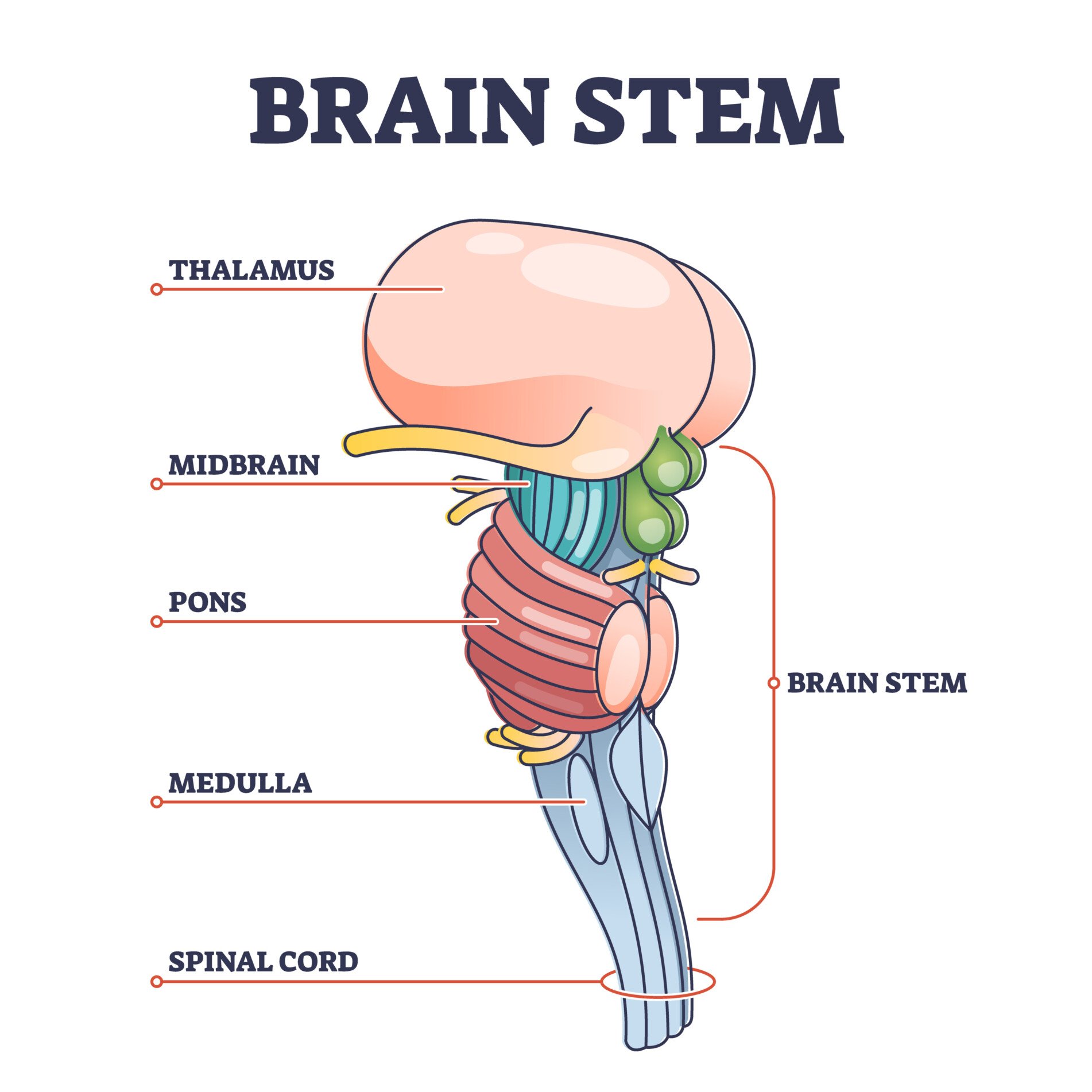
Brainstem
The brainstem is located at the base of the brain. This area connects the cerebrum and the cerebellum to the spinal cord, acting as a relay station for these areas.
The brainstem regulates automatic functions such as sleep cycles, breathing, body temperature, digestion, coughing, and sneezing.
Right Brain vs. Left Brain
The cerebrum is divided into two halves: theright and left hemispheres(Fig. 2). The left hemisphere controls the right half of the body, and the right hemisphere controls the left half.
The corpus callosum allows the two hemispheres to communicate and allows information being processed on one side of the brain to be shared with the other.

Figure 2.The cerebrum is divided into left and right hemispheres. The nerve fibers corpus callosum connects the two sides.
Hemispheric lateralizationis the idea that each hemisphere is responsible for different functions. Each of these functions is localized to either the right or left side.
The left hemisphere is associated with language functions, such as formulating grammar and vocabulary and containing different language centers (Broca’s and Wernicke’s area).
The right hemisphere is associated with more visuospatial functions such as visualization, depth perception, and spatial navigation. These left and right functions are the case in most people, especially those who are right-handed.
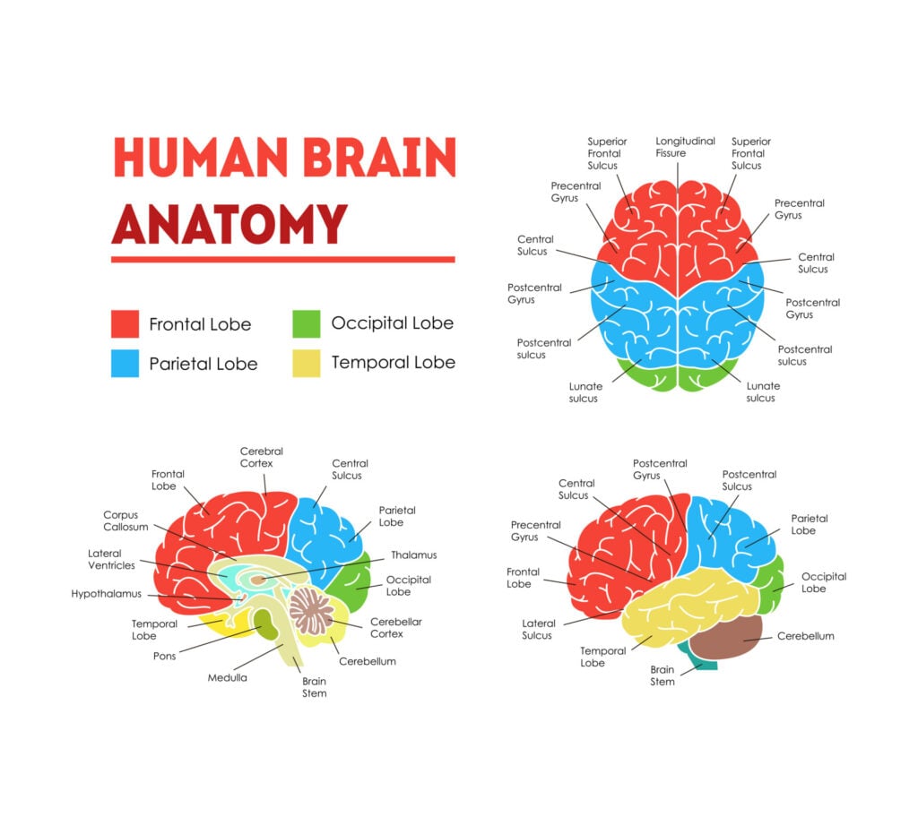
Lobes of the Brain
Each cerebral hemisphere can be subdivided into four lobes, each associated with different functions.
The four lobes of the brain are the frontal, parietal, temporal, and occipital lobes (Figure 3).

Figure 3.The cerebrum is divided into four lobes: frontal, parietal, occipital, and temporal.
Frontal lobesThefrontal lobesare located at the front of the brain, behind the forehead (Figure 4).Their main functions are associated with higher cognitive functions, including problem-solving, decision-making, attention, intelligence, and voluntary behaviors.The frontal lobes contain themotor cortexresponsible for planning and coordinating movements.It also contains the prefrontal cortex, which is responsible for initiating higher-lever cognitive functioning, and Broca’s Area, which is essential for language production.Figure 4.Frontal lobe structure.
Frontal lobesThefrontal lobesare located at the front of the brain, behind the forehead (Figure 4).Their main functions are associated with higher cognitive functions, including problem-solving, decision-making, attention, intelligence, and voluntary behaviors.
Frontal lobes
Thefrontal lobesare located at the front of the brain, behind the forehead (Figure 4).
Their main functions are associated with higher cognitive functions, including problem-solving, decision-making, attention, intelligence, and voluntary behaviors.
The frontal lobes contain themotor cortexresponsible for planning and coordinating movements.It also contains the prefrontal cortex, which is responsible for initiating higher-lever cognitive functioning, and Broca’s Area, which is essential for language production.Figure 4.Frontal lobe structure.
The frontal lobes contain themotor cortexresponsible for planning and coordinating movements.
It also contains the prefrontal cortex, which is responsible for initiating higher-lever cognitive functioning, and Broca’s Area, which is essential for language production.

Figure 4.Frontal lobe structure.
Temporal lobesThetemporal lobesare located on both sides of the brain, near the temples of the head, hence the name temporal lobes (Figure 5).The main functions of these lobes include understanding, language, memory acquisition, face recognition, object recognition, perception, and auditory information processing.There is a temporal lobe in both the left and right hemispheres. The left temporal lobe, which is usually the most dominant in people, is associated with language, learning, memorizing, forming words, and remembering verbal information.The left lobe also contains a vital language center known as Wernicke’s area, which is essential for language development. The right temporal lobe is usually associated with learning and memorizing non-verbal information and determining facial expressions.Figure 5.Temporal lobe structure.
Temporal lobes
Thetemporal lobesare located on both sides of the brain, near the temples of the head, hence the name temporal lobes (Figure 5).The main functions of these lobes include understanding, language, memory acquisition, face recognition, object recognition, perception, and auditory information processing.There is a temporal lobe in both the left and right hemispheres. The left temporal lobe, which is usually the most dominant in people, is associated with language, learning, memorizing, forming words, and remembering verbal information.The left lobe also contains a vital language center known as Wernicke’s area, which is essential for language development. The right temporal lobe is usually associated with learning and memorizing non-verbal information and determining facial expressions.Figure 5.Temporal lobe structure.
Thetemporal lobesare located on both sides of the brain, near the temples of the head, hence the name temporal lobes (Figure 5).
The main functions of these lobes include understanding, language, memory acquisition, face recognition, object recognition, perception, and auditory information processing.
There is a temporal lobe in both the left and right hemispheres. The left temporal lobe, which is usually the most dominant in people, is associated with language, learning, memorizing, forming words, and remembering verbal information.
The left lobe also contains a vital language center known as Wernicke’s area, which is essential for language development. The right temporal lobe is usually associated with learning and memorizing non-verbal information and determining facial expressions.

Figure 5.Temporal lobe structure.
Parietal lobesTheparietal lobeis located at the top of the brain, between the frontal and occipital lobes, and above the temporal lobes (Figure 6).The parietal lobe is essential for integrating information from the body’s senses to allow us to build a coherent picture of the world around us.These lobes allow us to perceive our bodies through somatosensory information (e.g., through touch, pressure, and temperature). It can also help with visuospatial processing, reading, and number representations (mathematics).The parietal lobes also contain the somatosensory cortex, which receives and processes sensory information, integrating this into a representational map of the body.This means it can pinpoint the exact area of the body where a sensation is felt, as well as perceive the weight of objects, shape, and texture.Figure 6.Parietal lobe structure.
Parietal lobes
Theparietal lobeis located at the top of the brain, between the frontal and occipital lobes, and above the temporal lobes (Figure 6).The parietal lobe is essential for integrating information from the body’s senses to allow us to build a coherent picture of the world around us.These lobes allow us to perceive our bodies through somatosensory information (e.g., through touch, pressure, and temperature). It can also help with visuospatial processing, reading, and number representations (mathematics).The parietal lobes also contain the somatosensory cortex, which receives and processes sensory information, integrating this into a representational map of the body.This means it can pinpoint the exact area of the body where a sensation is felt, as well as perceive the weight of objects, shape, and texture.Figure 6.Parietal lobe structure.
Theparietal lobeis located at the top of the brain, between the frontal and occipital lobes, and above the temporal lobes (Figure 6).
The parietal lobe is essential for integrating information from the body’s senses to allow us to build a coherent picture of the world around us.
These lobes allow us to perceive our bodies through somatosensory information (e.g., through touch, pressure, and temperature). It can also help with visuospatial processing, reading, and number representations (mathematics).
The parietal lobes also contain the somatosensory cortex, which receives and processes sensory information, integrating this into a representational map of the body.
This means it can pinpoint the exact area of the body where a sensation is felt, as well as perceive the weight of objects, shape, and texture.

Figure 6.Parietal lobe structure.
Occipital lobesTheoccipital lobesare located at the back of the brain behind the temporal and parietal lobes and below the occipital bone of the skull (Figure 7).The occipital lobes receive sensory information from the eyes’ retinas, which is then encoded into different visual data. Some of the functions of the occipital lobes include being able to assess the size, depth, and distance, determine color information, object and facial recognition, and mapping the visual world.The occipital lobes also contain the primary visual cortex, which receives sensory information from the retinas, transmitting this information relating to location, spatial data, motion, and the colors of objects in the field of vision.Figure 7.Occipital lobe structure.
Occipital lobes
Theoccipital lobesare located at the back of the brain behind the temporal and parietal lobes and below the occipital bone of the skull (Figure 7).The occipital lobes receive sensory information from the eyes’ retinas, which is then encoded into different visual data. Some of the functions of the occipital lobes include being able to assess the size, depth, and distance, determine color information, object and facial recognition, and mapping the visual world.The occipital lobes also contain the primary visual cortex, which receives sensory information from the retinas, transmitting this information relating to location, spatial data, motion, and the colors of objects in the field of vision.Figure 7.Occipital lobe structure.
Theoccipital lobesare located at the back of the brain behind the temporal and parietal lobes and below the occipital bone of the skull (Figure 7).
The occipital lobes receive sensory information from the eyes’ retinas, which is then encoded into different visual data. Some of the functions of the occipital lobes include being able to assess the size, depth, and distance, determine color information, object and facial recognition, and mapping the visual world.
The occipital lobes also contain the primary visual cortex, which receives sensory information from the retinas, transmitting this information relating to location, spatial data, motion, and the colors of objects in the field of vision.

Figure 7.Occipital lobe structure.
Cerebral Cortex
The surface of the cerebrum is called thecerebral cortexand has a wrinkled appearance, consisting of bulges, also known asgyri, and deep furrows, known as sulci(Figure 8).
A gyrus (plural: gyri) is the name given to the bumps and ridges on the cerebral cortex (the outermost layer of the brain). A sulcus (plural: sulci) is another name for a groove in the cerebral cortex.

The cerebral cortex is primarily constructed of grey matter (neural tissue made up of neurons), with between 14 and 16 billionneuronsfound here.
The many folds and wrinkles of the cerebral cortex allow a wider surface area for an increased number of neurons to live there, permitting large amounts of information to be processed.
Deep Structures
Amygdala
Theamygdalais a structure deep in the brain that is involved in the processing of emotions and fear learning. The amygdala is a part of the limbic system, a neural network that mediates emotion and memory (Figure 9).
This structure also ties emotional meaning to memories, processes rewards, and helps us make decisions. This structure has also been linked with the fight-or-flight response.

Thalamus and HypothalamusThethalamusrelays information between the cerebral cortex, brain stem, and other cortical structures (Figure 10).Because of its interactive role in relaying sensory and motor information, the thalamus contributes to many processes, including attention, perception, timing, and movement. The hypothalamus modulates a range of behavioral and physiological functions.It controls autonomic functions such as hunger, thirst, body temperature, and sexual activity. To do this, the hypothalamus integrates information from different brain parts and responds to various stimuli such as light, odor, and stress.Figure 10.The thalamus is often described as the brain’s relay station as a great deal of information that reaches the cerebral cortex first stops in the thalamus before being sent to its destination.
Thalamus and Hypothalamus
Thethalamusrelays information between the cerebral cortex, brain stem, and other cortical structures (Figure 10).Because of its interactive role in relaying sensory and motor information, the thalamus contributes to many processes, including attention, perception, timing, and movement. The hypothalamus modulates a range of behavioral and physiological functions.It controls autonomic functions such as hunger, thirst, body temperature, and sexual activity. To do this, the hypothalamus integrates information from different brain parts and responds to various stimuli such as light, odor, and stress.Figure 10.The thalamus is often described as the brain’s relay station as a great deal of information that reaches the cerebral cortex first stops in the thalamus before being sent to its destination.
Thethalamusrelays information between the cerebral cortex, brain stem, and other cortical structures (Figure 10).
Because of its interactive role in relaying sensory and motor information, the thalamus contributes to many processes, including attention, perception, timing, and movement. The hypothalamus modulates a range of behavioral and physiological functions.
It controls autonomic functions such as hunger, thirst, body temperature, and sexual activity. To do this, the hypothalamus integrates information from different brain parts and responds to various stimuli such as light, odor, and stress.

Figure 10.The thalamus is often described as the brain’s relay station as a great deal of information that reaches the cerebral cortex first stops in the thalamus before being sent to its destination.
HippocampusThehippocampusis a curved-shaped structure in the limbic system associated with learning and memory (Figure 11).This structure is most strongly associated with the formation of memories, is an early storage system for new long-term memories, and plays a role in the transition of these long-term memories to more permanent memories.Figure 11.Hippocampus location in the brain
Hippocampus
Thehippocampusis a curved-shaped structure in the limbic system associated with learning and memory (Figure 11).This structure is most strongly associated with the formation of memories, is an early storage system for new long-term memories, and plays a role in the transition of these long-term memories to more permanent memories.Figure 11.Hippocampus location in the brain
Thehippocampusis a curved-shaped structure in the limbic system associated with learning and memory (Figure 11).
This structure is most strongly associated with the formation of memories, is an early storage system for new long-term memories, and plays a role in the transition of these long-term memories to more permanent memories.

Figure 11.Hippocampus location in the brain
Basal GangliaThe basal ganglia are a group of structures that regulate the coordination of fine motor movements, balance, and posture alongside the cerebellum.These structures are connected to other motor areas and link the thalamus with the motor cortex. The basal ganglia are also involved in cognitive and emotional behaviors, as well as playing a role in reward and addiction.Figure 12.The Basal Ganglia Illustration
Basal Ganglia
The basal ganglia are a group of structures that regulate the coordination of fine motor movements, balance, and posture alongside the cerebellum.These structures are connected to other motor areas and link the thalamus with the motor cortex. The basal ganglia are also involved in cognitive and emotional behaviors, as well as playing a role in reward and addiction.Figure 12.The Basal Ganglia Illustration
The basal ganglia are a group of structures that regulate the coordination of fine motor movements, balance, and posture alongside the cerebellum.
These structures are connected to other motor areas and link the thalamus with the motor cortex. The basal ganglia are also involved in cognitive and emotional behaviors, as well as playing a role in reward and addiction.

Figure 12.The Basal Ganglia Illustration
Ventricles and Cerebrospinal Fluid
Within the brain, there are fluid-filled interconnected cavities calledventricles, which are extensions of the spinal cord. These are filled with a substance called cerebrospinal fluid, which is a clear and colorless liquid.
The ventricles produce cerebrospinal fluid and transport and remove this fluid. The ventricles do not have a unique function, but they provide cushioning to the brain and are useful for determining the locations of other brain structures.
Cerebrospinal fluid circulates the brain and spinal cord and functions to cushion the brain within the skull. If damage occurs to the skull, the cerebrospinal fluid will act as a shock absorber to help protect the brain from injury.

If there were a disruption or blockage, this can cause a build-up of cerebrospinal fluid and can cause enlarged ventricles.
Neurons
Neurons are thenerve cellsof the central nervous system that transmit information through electrochemical signals throughout the body. Neurons contain a soma, a cell body from which the axon extends.
Axons are nerve fibers that are the longest part of the neuron, which conduct electrical impulses away from the soma.
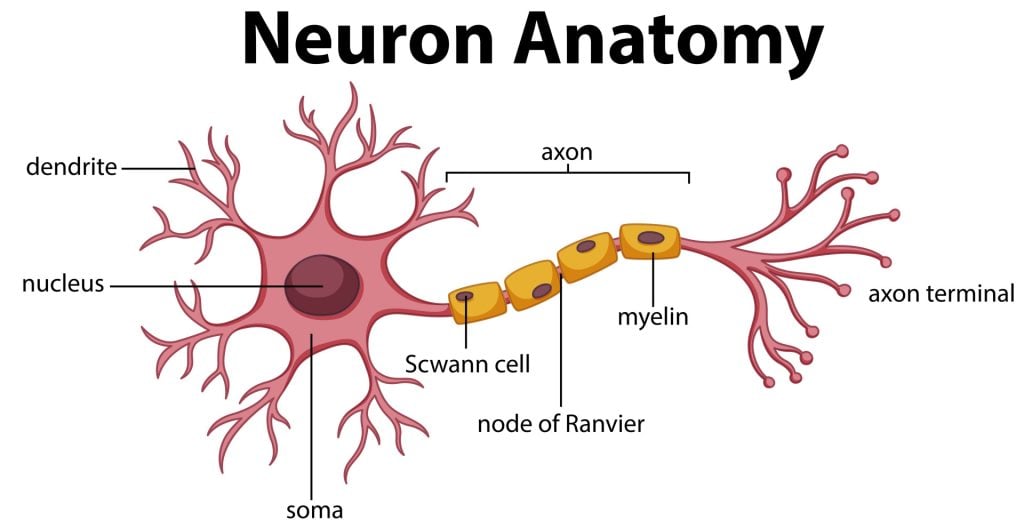
There are dendrites at the end of the neuron, which are branch-like structures that send and receive information from other neurons.
A myelin sheath, a fatty insulating layer, forms around the axon, allowing nerve impulses to travel down the axon quickly.
There are different types of neurons. Sensory neurons transmit sensory information, motor neurons transmit motor information, and relay neurons allow sensory and motor neurons to communicate.
The communication between neurons is called synapses. Neurons communicate with each other via synaptic clefts, which are gaps between the endings of neurons.

During synaptic transmission, chemicals, such as neurotransmitters, are released from the endings of the previous neuron (also known as the presynaptic neuron).
These chemicals enter the synaptic cleft to then be transported to receptors on the next neuron (also known as the postsynaptic neuron).
Once transported to the next neuron, the chemical messengers continue traveling down neurons to influence many functions, such as behavior and movement.
Glial Cells
Glial cellsare non-neuronal cells in the central nervous system which work to provide the neurons with nourishment, support, and protection.
Astrocytes
They also work to clean up what is left behind after synaptic transmission, either recycling any leftover neurotransmitters or cleaning up when a neuron dies.
Oligodendrocytes
This is a substance that is rich in fat and provides insulation to the neurons to aid neuronal signaling.
Microglial
Microglial cells have oval bodies and many branches projecting out of them. The primary function of these cells is to respond to injuries or diseases in the central nervous system.
Ependymal cellsThese cells are column-shaped and usually line up together to form a membrane called the ependyma. The ependyma is a thin membrane lining the spinal cord andventricles of the brain.In the ventricles, these cells have small hairlike structures called cilia, which help encourage the flow of cerebrospinal fluid.
Ependymal cells
These cells are column-shaped and usually line up together to form a membrane called the ependyma. The ependyma is a thin membrane lining the spinal cord andventricles of the brain.In the ventricles, these cells have small hairlike structures called cilia, which help encourage the flow of cerebrospinal fluid.
These cells are column-shaped and usually line up together to form a membrane called the ependyma. The ependyma is a thin membrane lining the spinal cord andventricles of the brain.
In the ventricles, these cells have small hairlike structures called cilia, which help encourage the flow of cerebrospinal fluid.
Cranial Nerves
There are12 types of cranial nerveswhich are linked directly to the brain without having to pass through the spinal cord. These allow sensory information to pass from the organs of the face to the brain:

Mnemonic for Order of Cranial Nerves:
SomeSayMarryMoneyButMyBrotherSaysBigBrainsMatterMore
References
Mayfield Brain and Spine (n.d.). Anatomy of the Brain. Retrieved July 28, 2021, from: https://mayfieldclinic.com/pe-anatbrain.htm
Robertson, S. (2018, August 23). What is Grey Matter? News Medical Life Sciences. https://www.news-medical.net/health/What-is-Grey-Matter.aspx
Guy-Evans, O. (2021, April 13). Temporal lobe: definition, functions, and location. Simply Psychology. www.www.www.www.www.www.simplypsychology.org/temporal-lobe.html
Guy-Evans, O. (2021, April 15). Parietal lobe: definition, functions, and location. Simply Psychology. www.www.www.www.www.www.simplypsychology.org/parietal-lobe.html
Guy-Evans, O. (2021, April 19). Occipital lobe: definition, functions, and location. Simply Psychology. www.www.www.www.www.www.simplypsychology.org/occipital-lobe.html
Guy-Evans, O. (2021, May 08). Frontal lobe function, location in brain, damage, more. Simply Psychology. www.www.www.www.www.www.simplypsychology.org/frontal-lobe.html
Guy-Evans, O. (2021, June 09). Gyri and sulci of the brain. Simply Psychology. www.www.www.www.www.www.simplypsychology.org/gyri-and-sulci-of-the-brain.html
 Human Brain Anatomy Infographic Card Poster System Concept of Diagnostics and Health Care Flat Design Style. Vector illustration of Head
 an illustration of the central nervous system with the lobes and areas labelled
![]()
Saul McLeod, PhD
BSc (Hons) Psychology, MRes, PhD, University of Manchester
Saul McLeod, PhD., is a qualified psychology teacher with over 18 years of experience in further and higher education. He has been published in peer-reviewed journals, including the Journal of Clinical Psychology.
Olivia Guy-Evans, MSc
BSc (Hons) Psychology, MSc Psychology of Education
Olivia Guy-Evans is a writer and associate editor for Simply Psychology. She has previously worked in healthcare and educational sectors.
