On This Page:ToggleFunctionsLocationSubstructuresDamage
On This Page:Toggle
On This Page:
The parietal lobe,located in the upper middle part of the cerebral cortex, plays a central role in integrating sensory information from various body parts, understanding spatial orientation, and processing information about touch.

What does the parietal lobe do?
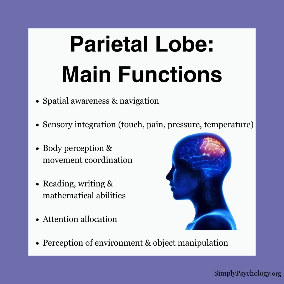
The parietal lobe plays a crucial role inintegrating sensory informationfrom various modalities and is associated with several key functions:
Spatial Awareness and Navigation
Sensory Integration and Processing
Language and Mathematics
Where is the parietal lobe located?
The brain’s parietal lobe is situatedbetween the frontal andoccipital lobesand above thetemporal lobes. The parietal lobes take up premises in the brain’s right and left hemispheres.
It makes up about19% of the brain’s cerebral cortexand is made up of neurons that send or relay signals to other neurons, andglial cells, that support the neurons.
The parietal lobes play a crucial role in processing sensory information, spatial awareness, perception, attention, and the integration of sensory inputs with motor functions.

Substructures of the Parietal Lobe
The parietal lobe is structurally divided into thesomatosensory cortex, inferior parietal lobe, superior parietal lobe, and precuneus.
Somatosensory Cortex
Thesomatosensory cortex’smain function is to receive and process sensory information from the entire body, such as touch, temperature, and pain.
It creates arepresentational map of the bodywithin the brain and is responsible for localizing sensations, perceiving different degrees of pressure, and recognizing the shape and texture of objects through touch.
Inferior Parietal Lobe
The inferior parietal lobe is primarily concerned with language, mathematical operations, and body image.
It is also important forspatial attention, visuomotor, and auditory processing, and has been suggested to be involved in the perception of emotions through facial expressions.
Damage to this area may result in impairments inspeech repetitionand difficulty completing mathematical problems.
Superior Parietal Lobe
The superior parietal lobe is concerned withspatial orientation and sensorimotor integration, and it receives a lot of visual and sensory signals from the hands.
Precuneus
The precuneus is located on the medial surface of the parietal lobes and is one of the least accurately mapped areas of the cortex.
Functional neuroimaging suggests that the precuneus is involved in tasks such asvisuospatial imagery,episodic memory retrieval, and theability to take first-person perspectives.
This area is also thought to play a role in self-awareness and consciousness.

What Happens When the Parietal Lobe is Damaged?
Damage to the parietal lobes, which can result from conditions like stroke, vascular disease, tumors, traumatic brain injury, or infections, can lead to a variety of symptoms:
Research has provided further insights into parietal lobe damage:
Overall, parietal lobe damage cansignificantly impact an individual’s perception, sensory integration, spatial awareness, language, and cognitive abilities, with distinct deficits arising from left versus right-side lesions. Ongoing research continues to elucidate the complex functions of this brain region.
References
Cavanna, A. E., & Trimble, M. R. (2006). The precuneus: a review of its functional anatomy and behavioural correlates.Brain,129(3), 564-583.
Ferro, A., Bonivento, C., Delvecchio, G., Bellani, M., Perlini, C., Dusi, N., Marinelli, V., Ruggeri, M., Altamura, C., Crespo-Facorro. & Brambilla, P. (2017). Longitudinal investigation of the parietal lobe anatomy in bipolar disorder and its association with general functioning. Psychiatry Research: Neuroimaging, 267, 22-31.
Freund, H. J. (2003). Somatosensory and motor disturbances in patients with parietal lobe lesions.Advances in Neurology, 93, 179-193.
Fridriksson, J., Kjartansson, O., Morgan, P. S., Hjaltason, H., Magnusdottir, S., Bonilha, L., & Rorden, C. (2010). Impaired speech repetition and left parietal lobe damage. Journal of Neuroscience, 30(33), 11057-11061.
Milner, A. D. (1998). Streams and consciousness: visual awareness and the brain.Trends in Cognitive Sciences, 2(1), 25-30.
Radua, J., Phillips, M. L., Russell, T., Lawrence, N., Marshall, N., Kalidindi, S., El-Hage, W., McDonald, C., Giampietro, V., Brammer, David, A. S. & Surguladze, S. A. (2010). Neural response to specific components of fearful faces in healthy and schizophrenic adults.Neuroimage, 49(1), 939-946.
Torrey, E. F. (2007). Schizophrenia and the inferior parietal lobule.Schizophrenia Research, 97(1-3), 215-225.
Vance, A., Silk, T. J., Casey, M., Rinehart, N. J., Bradshaw, J. L., Bellgrove, M. A., & Cunnington, R. (2007). Right parietal dysfunction in children with attention deficit hyperactivity disorder, combined type: a functional MRI study.Molecular Psychiatry, 12(9), 826-832.
Weiskrantz, L. (1977). Trying to bridge some neuropsychological gaps between monkey and man.British Journal of Psychology, 68(4), 431-445.
Zhou, S. Y., Suzuki, M., Takahashi, T., Hagino, H., Kawasaki, Y., Matsui, M., Seto, H. & Kurachi, M. (2007). Parietal lobe volume deficits in schizophrenia spectrum disorders.Schizophrenia Research, 89(1-3), 35-48.
![]()
Saul McLeod, PhD
BSc (Hons) Psychology, MRes, PhD, University of Manchester
Saul McLeod, PhD., is a qualified psychology teacher with over 18 years of experience in further and higher education. He has been published in peer-reviewed journals, including the Journal of Clinical Psychology.
Olivia Guy-Evans, MSc
BSc (Hons) Psychology, MSc Psychology of Education
Olivia Guy-Evans is a writer and associate editor for Simply Psychology. She has previously worked in healthcare and educational sectors.
