On This Page:ToggleFunctionsLocationStructureDamage
On This Page:Toggle
On This Page:
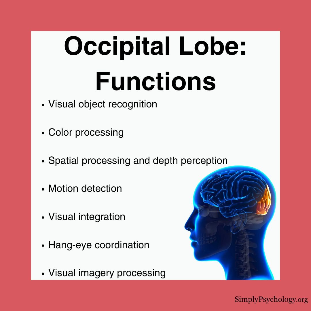
The occipital lobesare a part of the brain responsible for processing visual information, such as object recognition, color perception, depth perception, and motor detection.
Damage to these lobes can result invisual impairments, such as difficulty recognizing objects or disturbances in visual perception.
Through a complex network of connections with other brain regions, the occipital lobes contribute to our ability to navigate the visual world, form mental images, and perceive the surrounding environment.

Functions
The occipital lobeprocesses all visual informationthrough several specialized systems:
Visual Recognition
Spatial Processing
Color Processing
Integration with Other Brain Regions
Unique Visual Phenomena
The occipital lobe is involved in some fascinating visual experiences:
Location
The occipital lobe is located at the very back of the brain beneath the occipital bone andbehind theparietalandtemporal lobes.
It is the smallest lobe of the brain, accounting for around12% of the total surface area of the brain’s cortex.
The occipital lobe, located at the back of the brain, is primarily responsible for visual processing. It interprets visual information received from the eyes, enabling us to recognize and understand what we see. Damage to this lobe can lead to visual deficits or disturbances.

Structure
The occipital lobes can bedivided into several functional areas, although no anatomical markers distinguish these areas.

Primary Visual Cortex
This section is also known as Brodmann area 17, or visual area V1. The primary visual cortex receivessensory information from the eyes’ retinasand then transmits information relating to location, spatial data, motion, and the colors of objects in the field of vision.
This information gets transported via two streams: the dorsal and ventral streams. The visual cortex is divided into six areas depending on the function and structure of each area, referred to as V1, V2, V3, V4, and V5.
Secondary Visual Cortex
This section is also known asBrodmann area 18 and 19, or visual area V2. This is the area immediately surrounding the primary visual cortex.
It receives information from the primary visual cortex for furtherorganization of visual input. It also passes information to visual areas V3, V4, and V5.
Ventral Stream
The secondary visual cortex also encompasses the ventral stream, whichallows information to flow to temporal lobestructures to enable us to process what objects are.
Without the ventral stream, we would still be able to see normally without conscious awareness or understanding of what we see.
Lateral Geniculate Bodies
The lateral geniculate body is part of thethalamusand is asensory relay system.
Raw information coming from the outer part of the retinas enters this area for processing before being sent to the primary visual cortex.
Lingula
With the help of the lateral geniculate bodies, the lingula creates spatial awareness and gives depth to visual information.
Dorsal Stream
The dorsal stream allows information to flow from the occipital lobes to the parietal lobes in order for us toprocess where objects are located.
The dorsal stream connects to both the V1 and V2 regions, allowing these areas to send information about the size and shape of objects in our field of vision.
Damage and associated conditions
Below are some signs that may indicate damage to the occipital lobes:
How Damage Occurs
The occipital lobe’s position at the back of the brainprovides some natural protection from injury. However, damage can occur through trauma, stroke, tumors, or infections.
Interestingly, if only part of the primary visual cortex is damaged, it can create a specific “blind spot” in the person’s field of vision, corresponding to the damaged area.
Associated Conditions
Studies using MRIhave revealed significant occipital cortex enlargement inautistic individualscompared to control groups.
This structural difference correlates withenhanced perceptual abilities, particularly in visual search and discrimination tasks, where individuals with autismoften outperform neurotypical participants.
Research using PET scanshas identified a correlation between depression and reduced blood flow in the occipital lobe.
They found that successful pharmacotherapy treatment for depression can increase blood flow to the parieto-occipital regions, suggesting a previously unknownlink between visual processing and mood disorders.
Occipital lobe epilepsy presents unique challenges for treatment. While surgery can be successful, precise identification of the affected area through brain imaging remains difficult.
Studies have shownthat individuals with occipital lobe epilepsy oftendemonstrate reduced performance in attention, memory, and intellectual functioning compared to control groups.
Brain imaging studieshave revealed reducedgray mattervolume in the visual association cortex of individuals with chronic schizophrenia.
This structural difference may explain some of thevisual processing deficits commonly observed in schizophrenia patients.
The connection between occipital lobe abnormalities and visual hallucinations in schizophrenia continues to be an active area of research.
ReferencesBinder, D. K., Von Lehe, M., Kral, T., Bien, C. G., Urbach, H., Schramm, J., & Clusmann, H. (2008). Surgical treatment of occipital lobe epilepsy.Journal of Neurosurgery, 109(1), 57-69.Gülgönen, S., Demirbilek, V., Korkmaz, B., Dervent, A., & Townes, B. D. (2000). Neuropsychological functions in idiopathic occipital lobe epilepsy.Epilepsia, 41(4), 405-411.Harward, S. C., Chen, W. C., Rolston, J. D., Haglund, M. M., & Englot, D. J. (2018). Seizure outcomes in occipital lobe and posterior quadrant epilepsy surgery: a systematic review and meta-analysis.Neurosurgery, 82(3), 350-358.Ishizaki, J., Yamamoto, H., Takahashi, T., Takeda, M., Yano, M., & Mimura, M. (2008). Changes in regional cerebral blood flow following antidepressant treatment in late‐life depression.International Journal of Geriatric Psychiatry, 23(8), 805-811.Jessell, T. M. (1991).Principles of neural science(pp. 173-193). J. H. Schwartz, & E. R. Kandel (Eds.). New York: Elsevier.McCarley, R. W., Wible, C. G., Frumin, M., Hirayasu, Y., Levitt, J. J., Fischer, I. A., & Shenton, M. E. (1999). MRI anatomy of schizophrenia.Biological Psychiatry, 45(9), 1099-1119.Onitsuka, T., McCarley, R. W., Kuroki, N., Dickey, C. C., Kubicki, M., Demeo, S. S., Frumin, M., Kikinis, R., Jolesz, F. A. & Shenton, M. E. (2007). Occipital lobe gray matter volume in male patients with chronic schizophrenia: A quantitative MRI study.Schizophrenia Research, 92(1-3), 197-206.Park, K. C., Yoon, S. S., & Rhee, H. Y. (2011). Executive dysfunction associated with stroke in the posterior cerebral artery territory.Journal of Clinical Neuroscience, 18(2), 203-208.Piven, J., Arndt, S., Bailey, J., & Andreasen, N. (1996). Regional brain enlargement in autism: a magnetic resonance imaging study.Journal of the American Academy of Child & Adolescent Psychiatry, 35(4), 530-536.Samson, F., Mottron, L., Soulières, I., & Zeffiro, T. A. (2012). Enhanced visual functioning in autism: An ALE meta‐analysis.Human Brain Mapping, 33(7), 1553-1581.Sperling, J. M., Prvulovic, D., Linden, D. E., Singer, W., & Stirn, A. (2006). Neuronal correlates of colour-graphemic synaesthesia: AfMRI study.Cortex, 42(2), 295-303.Tate, D. F., Bigler, E. D., McMahon, W., & Lainhart, J. (2007). The relative contributions of brain, cerebrospinal fluid-filled structures and non-neural tissue volumes to occipital-frontal head circumference in subjects with autism. Neuropediatrics, 38(01), 18-24.
References
Binder, D. K., Von Lehe, M., Kral, T., Bien, C. G., Urbach, H., Schramm, J., & Clusmann, H. (2008). Surgical treatment of occipital lobe epilepsy.Journal of Neurosurgery, 109(1), 57-69.
Gülgönen, S., Demirbilek, V., Korkmaz, B., Dervent, A., & Townes, B. D. (2000). Neuropsychological functions in idiopathic occipital lobe epilepsy.Epilepsia, 41(4), 405-411.
Harward, S. C., Chen, W. C., Rolston, J. D., Haglund, M. M., & Englot, D. J. (2018). Seizure outcomes in occipital lobe and posterior quadrant epilepsy surgery: a systematic review and meta-analysis.Neurosurgery, 82(3), 350-358.
Ishizaki, J., Yamamoto, H., Takahashi, T., Takeda, M., Yano, M., & Mimura, M. (2008). Changes in regional cerebral blood flow following antidepressant treatment in late‐life depression.International Journal of Geriatric Psychiatry, 23(8), 805-811.
Jessell, T. M. (1991).Principles of neural science(pp. 173-193). J. H. Schwartz, & E. R. Kandel (Eds.). New York: Elsevier.
McCarley, R. W., Wible, C. G., Frumin, M., Hirayasu, Y., Levitt, J. J., Fischer, I. A., & Shenton, M. E. (1999). MRI anatomy of schizophrenia.Biological Psychiatry, 45(9), 1099-1119.
Onitsuka, T., McCarley, R. W., Kuroki, N., Dickey, C. C., Kubicki, M., Demeo, S. S., Frumin, M., Kikinis, R., Jolesz, F. A. & Shenton, M. E. (2007). Occipital lobe gray matter volume in male patients with chronic schizophrenia: A quantitative MRI study.Schizophrenia Research, 92(1-3), 197-206.
Park, K. C., Yoon, S. S., & Rhee, H. Y. (2011). Executive dysfunction associated with stroke in the posterior cerebral artery territory.Journal of Clinical Neuroscience, 18(2), 203-208.
Piven, J., Arndt, S., Bailey, J., & Andreasen, N. (1996). Regional brain enlargement in autism: a magnetic resonance imaging study.Journal of the American Academy of Child & Adolescent Psychiatry, 35(4), 530-536.
Samson, F., Mottron, L., Soulières, I., & Zeffiro, T. A. (2012). Enhanced visual functioning in autism: An ALE meta‐analysis.Human Brain Mapping, 33(7), 1553-1581.
Sperling, J. M., Prvulovic, D., Linden, D. E., Singer, W., & Stirn, A. (2006). Neuronal correlates of colour-graphemic synaesthesia: AfMRI study.Cortex, 42(2), 295-303.
Tate, D. F., Bigler, E. D., McMahon, W., & Lainhart, J. (2007). The relative contributions of brain, cerebrospinal fluid-filled structures and non-neural tissue volumes to occipital-frontal head circumference in subjects with autism. Neuropediatrics, 38(01), 18-24.
![]()
Saul McLeod, PhD
BSc (Hons) Psychology, MRes, PhD, University of Manchester
Saul McLeod, PhD., is a qualified psychology teacher with over 18 years of experience in further and higher education. He has been published in peer-reviewed journals, including the Journal of Clinical Psychology.
Olivia Guy-Evans, MSc
BSc (Hons) Psychology, MSc Psychology of Education
Olivia Guy-Evans is a writer and associate editor for Simply Psychology. She has previously worked in healthcare and educational sectors.
